
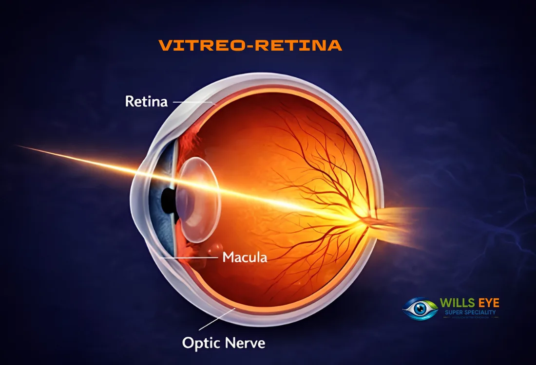
Retina Clinic & Surgery
The retina is the light-sensing layer lining the back of the eye, and retinal treatment addresses conditions such as tears, detachment, bleeding and swelling that can cause permanent vision loss if untreated.
Speak to a specialist about Retina Clinic & Surgery in Agartala.
What is Retina Clinic & Surgery?
The retina is the light-sensing layer lining the back of the eye, and retinal treatment addresses conditions such as tears, detachment, bleeding and swelling that can cause permanent vision loss if untreated.
Also known as: Vitreo-retinal services · Retina treatment · Fundus clinic
Retinal disease is quiet until it is not. Much of the damage happens without pain and often without the patient noticing, which is why a dilated fundus examination is part of every complete eye check-up we do.
Request an appointment
Talk to our specialist about Retina Clinic & Surgery.
Request Appointment Call Us on 8583948238 or message on WhatsApp- Location
- Colonel Chowmuhani, Agartala
- Serving
- All 8 districts of Tripura
- Cashless
- TPA & insurance
Retinal warning signs — see us the same day
- A sudden shower of new floaters, black spots or “soot” across your vision
- Flashes of light, especially in a dark room or when moving your eyes
- A dark curtain or shadow spreading in from one side of your field of vision
- Straight lines — door frames, tiles, text — appearing bent or wavy
- A blurred or missing patch in the centre of what you are looking at
- Sudden painless loss of vision in one eye
Conditions treated in the retina clinic
- Diabetic retinopathy and diabetic macular oedema
- Age-related macular degeneration, both dry and wet types
- Retinal tears, holes and retinal detachment
- Retinal vein and artery occlusions
- Vitreous haemorrhage — bleeding into the gel cavity of the eye
- Myopic retinal degeneration in highly short-sighted eyes
Treatment options at Wills Eye
Dilated fundus evaluation and OCT
Indirect ophthalmoscopy plus optical coherence tomography, which images the retinal layers in cross-section and detects swelling long before vision drops.
Retinal laser photocoagulation
Out-patient laser used to seal tears, treat leaking vessels and stabilise diabetic retinopathy.
Intravitreal injections
Anti-VEGF and steroid injections for macular swelling and abnormal new vessels.
Vitreo-retinal surgery
Vitrectomy and detachment repair for advanced disease, with referral pathways where a specific tertiary intervention is required.
What to expect — step by step
-
1
Dilation
Drops widen the pupil so the entire retina, including the far periphery, can be examined. Vision stays blurred for four to six hours.
-
2
Imaging
Fundus photography and OCT to document the baseline and to compare accurately at future visits.
-
3
Diagnosis and plan
The findings are explained with the images in front of you, along with what will happen if treatment is deferred.
-
4
Treatment and monitoring
Laser, injection or surgery as indicated, then a defined review interval — retinal disease is managed over time, not in a single visit.
Recovery & aftercare
Recovery depends entirely on the procedure. Laser is out-patient with mild ache for a day. Injections need a few days of care against infection. Vitrectomy and detachment repair involve a longer course, sometimes including posturing. What is common to all is that follow-up is not optional — retinal conditions are monitored, not simply fixed and forgotten.
Why timing matters
Retinal tissue is nerve tissue. Unlike a cornea or a lens, it does not regenerate, so vision already lost to a detached or scarred retina cannot be recovered — only what remains can be protected. That is the entire argument for coming in on the day you notice flashes, floaters or a shadow, rather than waiting to see if it settles.
Retina Clinic & Surgery — frequently asked questions
Are floaters always serious? +
Long-standing, unchanged floaters are usually harmless. A sudden increase, especially with flashes or a shadow, needs a dilated examination the same day because it can mean a retinal tear.
Will my vision come back after retinal treatment? +
Treatment aims first to stop further loss. Some vision often recovers, particularly when swelling is treated early, but retina that has already been destroyed cannot be restored.
How often should a diabetic have a retina check? +
At least once a year from the time of diagnosis, and more frequently if any retinopathy is already present or during pregnancy.
Is dilation necessary every time? +
For a proper retinal assessment, yes. The peripheral retina — where tears and detachments usually begin — simply cannot be seen through an undilated pupil.
Meet our eye specialists
Consultations for retina clinic & surgery are with the ophthalmologists at Wills Eye, Agartala.
- Dr. Partha Pratim Pal — Founder & Chief Ophthalmologist
Travelling from outside Agartala?
Patients come to us from every district of Tripura. Find the route and travel time from your town under eye care across Tripura, see how cashless treatment works, how treatment charges are worked out, or read the frequently asked questions.
This page is written for general information about retina clinic & surgery and is not a substitute for examination and advice from a qualified eye doctor. Treatment recommendations depend on your individual findings.